Saturday, November 29, 2008
The following observations refer specifically to the tank labeled with the following code, top to bottom: Red C, Red L, Blue P, stored in laboratory section 004, Wednesday 11:15. This tank includes pond water from Container # 1 (Tommy Schumpert Pond, Seven Islands Wildlife Refuge, Kelly Lane, Knox Co. Tennessee. Partial shade exposure. Sheet runoff around sink hole. N35 57.256 W83 41.503 947 10/12/2008) with some existing algal plant life, along with Plant B (Utricularia vulgaris L. A flowering, carnivorous plant. Collection from: Greenhouse in White Avenue Biology Annex. The University of Tennessee. 1400 White Ave. Knox Co. Knoxville TN. Partial shade exposure N 35o57' 33.45" W083o55' 42.01". 932 ft 10/13/2008). The associated Blog for this tank, noting more complete observations is available online at <http://microcosmicgod.blogspot.com/ >.
Upon initial observation immediately following tank setup on 15 October 2008, the water appeared quite clouded to the unaided eye, but a wide variety of living organisms were evident under 10X magnification in the binocular compound light microscope. Especially notable upon this first observation were many small flagellates, ciliates and rotifers throughout the tank. Also noted were several apparent insect larvae and one large egg-bearing female Macrocyclops near the plant Utricularia vulgaris, the common bladderwort identified by several bladder-shaped green leaves.
One week later, on 22 October 2008, the tank had lost at least 30% of the original moisture, which was replaced by dropper with distilled water. This observation yielded several desmids, identified by the greenish colors, distinctive rod or “cucumber” shapes and the nuclei at restricta (<http://www.microscopy-uk.org.uk/index.html?http://www.microscopy-uk.org.uk/pond/index.html>). Another Macrocyclops was sighted, identified as an adult male due to the fully developed appendages and lack of egg sacs (). The Macrocyclops albidus is a copepod crustacean and is discussed in further detail in the following section. Several algae were also noted upon this observation but had not yet been classified.
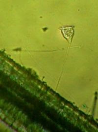
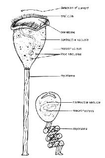
The third observation on 29 October 2008 occurred five days after a single pellet of Atison’s Betta Food had been added to the tank (McFarland). Most of the previously noted organisms were still present, but several new species were also evident. Of particular note was the ciliated protist Vorticella, several of which were attached by stalks to the stems of Utricularia (Corliss 273). When disrupted by movement, the bell-shaped body of the organism closes quickly and a structure called the myoneme coils the stalk like a spring, making the organism appear much smaller, presumably as a deterrent to predators (<http://www.microscope-microscope.org/applications/pond-critters/protozoans/ciliphora/vorticella.htm>). Also noted were a large number of rapidly moving Paramecium ciliates identified by their cigar shape and peristomata (), as well as a substantial number of new growth crosiers on the carnivorous plant Utricularia.
The following week, on 7 November 2008, a variety of diatoms were noted near the bottom of the tank, identified by their golden and green color and strikingly symmetrical shapes (Raven 275). Also noted was the emergence of a Chlorophyte alga, branching prolifically throughout the tank (Raven 391). In addition, several small areas included Spirogyra, identified by the unique spiral shape of the chloroplasts in thin branching filaments (Forest 89).
On the final day of observation, 14 November 2008, a notable change in plant life had occurred. While Utricularia remained vibrant, the alga Stigeoclonium nanum had become prolific throughout the tank, and many small rotifers and ciliates seemed attracted to its “branches” (Forest 87). Meanwhile, the now plentiful Macrocyclops seemed to favor the protection or environment of Utricularia (both discussed in detail in the following section).
Classification and Results
Special consideration was given in this study to two microorganisms that commonly coexist in nature. Utricularia macrorhiza, the common Bladderwort, and Macrocyclops albidus, the copepod commonly known as Cyclops, are both abundant in freshwater ponds in the southeastern United States, with the Bladderwort regularly supplying shelter for Cyclops (http://www.fcps.edu/islandcreekes/ecology/copepod.html).
Both organisms regularly prey on insect larvae, especially mosquitoes, and have been the subjects of several studies involving their possible uses as natural pesticides in mosquito control in tropical and sub-tropical climates. The Blog associated with the classroom study in this project (<http://microcosmicgod.blogspot.com/ >) includes references and links to an ongoing study at the University of Florida, in which the copepods are cultivated in used tires filled with stagnant water and then studied for their effects on mosquito populations (<http://fmel.ifas.ufl.edu/COPEPODS.HTM>). The Bladderwort, a carnivorous flowering aquatic plant, has also been studied for its possible use in mosquito control, but concern remains among scientists that Utricularia itself may in fact become an uncontrollable genus if intentionally cultivated in areas rich in mosquito larval food sources, because of its widespread rhizomes its and ability to asexually reproduce by fragmentation (Taylor 182). Following are brief discussions about both of these organisms, including taxonomy, life cycles and descriptions. Additional photographs and graphic displays are posted and their sources cited at (<http://microcosmicgod.blogspot.com/ >).
Utricularia vulgaris L.
The taxonomy of Utricularia is somewhat vague, with some dissention among scientists as to the species represented here. In general, however, Utricularia vulgaris and Utricularia macrorhiza are so similar in most respects that they may be considered together for the purposes of this study (Figure 1) ().
DOMAIN
Eukarya
KINGDOM
Plantae, the Plants
PHYLUM
Magnoliophyta (Angiosperms, the Flowering Plants)
CLASS
Magnoliopsida, the Dicotyledons
Subclass Asteridae
ORDER
Scrophulariales
FAMILY
Lentibulariaceae – Bladderwort family
GENUS
Utricularia
SPECIES
Utricularia macrorhiza, Utricularia vulgaris
Figure 1 Utricularia Classification
Collected from the greenhouse at the White Avenue Biology Annex, this carnivorous flowering plant is also known as the common Bladderwort. The plant consists of a network of underwater stems called rhizomes equipped with oval, bladder-shaped leaves (Figures 3, 4). Above the water’s surface, Utricularia produces delicate yellow flowers similar in appearance to snapdragon flowers, and reproduces sexually as a dicot angiosperm (<www.rook.org/earl/bwca/nature/aquatics/utriculariamac.html>). The bladders, long thought to provide floatation for the underwater portions of the plant, actually act as traps for small prey. When the tiny, cilia-like hairs at the bladder opening sense motion, the bladder closes, trapping inside any insect larvae or other small organisms with the misfortune of swimming past. Once the prey is inside, Utricularia secretes enzymes to slowly break down the matter into forms which can be ingested and used as nutrition for the plant. Thus, this carnivorous plant is able to gain nutrients not only through photosynthesis but also through this complicated digestive process. Utricularia reproduces sexually as an Angiosperm (Figure 2), and is additionally able to reproduce asexually through fragmentation.
Figure 2 Life Cycle of Utricularia (Adapted from Angiosperm Life Cycle by Sarah Pena)
Upon initial observation immediately following tank setup on 15 October 2008, the water appeared quite clouded to the unaided eye, but a wide variety of living organisms were evident under 10X magnification in the binocular compound light microscope. Especially notable upon this first observation were many small flagellates, ciliates and rotifers throughout the tank. Also noted were several apparent insect larvae and one large egg-bearing female Macrocyclops near the plant Utricularia vulgaris, the common bladderwort identified by several bladder-shaped green leaves.
One week later, on 22 October 2008, the tank had lost at least 30% of the original moisture, which was replaced by dropper with distilled water. This observation yielded several desmids, identified by the greenish colors, distinctive rod or “cucumber” shapes and the nuclei at restricta (<http://www.microscopy-uk.org.uk/index.html?http://www.microscopy-uk.org.uk/pond/index.html>). Another Macrocyclops was sighted, identified as an adult male due to the fully developed appendages and lack of egg sacs (
The third observation on 29 October 2008 occurred five days after a single pellet of Atison’s Betta Food had been added to the tank (McFarland). Most of the previously noted organisms were still present, but several new species were also evident. Of particular note was the ciliated protist Vorticella, several of which were attached by stalks to the stems of Utricularia (Corliss 273). When disrupted by movement, the bell-shaped body of the organism closes quickly and a structure called the myoneme coils the stalk like a spring, making the organism appear much smaller, presumably as a deterrent to predators (<http://www.microscope-microscope.org/applications/pond-critters/protozoans/ciliphora/vorticella.htm>). Also noted were a large number of rapidly moving Paramecium ciliates identified by their cigar shape and peristomata (
The following week, on 7 November 2008, a variety of diatoms were noted near the bottom of the tank, identified by their golden and green color and strikingly symmetrical shapes (Raven 275). Also noted was the emergence of a Chlorophyte alga, branching prolifically throughout the tank (Raven 391). In addition, several small areas included Spirogyra, identified by the unique spiral shape of the chloroplasts in thin branching filaments (Forest 89).
On the final day of observation, 14 November 2008, a notable change in plant life had occurred. While Utricularia remained vibrant, the alga Stigeoclonium nanum had become prolific throughout the tank, and many small rotifers and ciliates seemed attracted to its “branches” (Forest 87). Meanwhile, the now plentiful Macrocyclops seemed to favor the protection or environment of Utricularia (both discussed in detail in the following section).
Classification and Results
Special consideration was given in this study to two microorganisms that commonly coexist in nature. Utricularia macrorhiza, the common Bladderwort, and Macrocyclops albidus, the copepod commonly known as Cyclops, are both abundant in freshwater ponds in the southeastern United States, with the Bladderwort regularly supplying shelter for Cyclops (http://www.fcps.edu/islandcreekes/ecology/copepod.html).
Both organisms regularly prey on insect larvae, especially mosquitoes, and have been the subjects of several studies involving their possible uses as natural pesticides in mosquito control in tropical and sub-tropical climates. The Blog associated with the classroom study in this project (<http://microcosmicgod.blogspot.com/ >) includes references and links to an ongoing study at the University of Florida, in which the copepods are cultivated in used tires filled with stagnant water and then studied for their effects on mosquito populations (<http://fmel.ifas.ufl.edu/COPEPODS.HTM>). The Bladderwort, a carnivorous flowering aquatic plant, has also been studied for its possible use in mosquito control, but concern remains among scientists that Utricularia itself may in fact become an uncontrollable genus if intentionally cultivated in areas rich in mosquito larval food sources, because of its widespread rhizomes its and ability to asexually reproduce by fragmentation (Taylor 182). Following are brief discussions about both of these organisms, including taxonomy, life cycles and descriptions. Additional photographs and graphic displays are posted and their sources cited at (<http://microcosmicgod.blogspot.com/ >).
Utricularia vulgaris L.
The taxonomy of Utricularia is somewhat vague, with some dissention among scientists as to the species represented here. In general, however, Utricularia vulgaris and Utricularia macrorhiza are so similar in most respects that they may be considered together for the purposes of this study (Figure 1) (
DOMAIN
Eukarya
KINGDOM
Plantae, the Plants
PHYLUM
Magnoliophyta (Angiosperms, the Flowering Plants)
CLASS
Magnoliopsida, the Dicotyledons
Subclass Asteridae
ORDER
Scrophulariales
FAMILY
Lentibulariaceae – Bladderwort family
GENUS
Utricularia
SPECIES
Utricularia macrorhiza, Utricularia vulgaris
Figure 1 Utricularia Classification
Collected from the greenhouse at the White Avenue Biology Annex, this carnivorous flowering plant is also known as the common Bladderwort. The plant consists of a network of underwater stems called rhizomes equipped with oval, bladder-shaped leaves (Figures 3, 4). Above the water’s surface, Utricularia produces delicate yellow flowers similar in appearance to snapdragon flowers, and reproduces sexually as a dicot angiosperm (<www.rook.org/earl/bwca/nature/aquatics/utriculariamac.html>). The bladders, long thought to provide floatation for the underwater portions of the plant, actually act as traps for small prey. When the tiny, cilia-like hairs at the bladder opening sense motion, the bladder closes, trapping inside any insect larvae or other small organisms with the misfortune of swimming past. Once the prey is inside, Utricularia secretes enzymes to slowly break down the matter into forms which can be ingested and used as nutrition for the plant. Thus, this carnivorous plant is able to gain nutrients not only through photosynthesis but also through this complicated digestive process. Utricularia reproduces sexually as an Angiosperm (Figure 2), and is additionally able to reproduce asexually through fragmentation.
Figure 2 Life Cycle of Utricularia (Adapted from Angiosperm Life Cycle by Sarah Pena)
Saturday, November 15, 2008
Utricularia notes
USDA Forest Service May 2006
www.fs.fed.us/r2/projects/scp/assessments/utriculariaminor.pdf
population/community ecology
www.fs.fed.us/r2/projects/scp/assessments/utriculariaminor.pdf
population/community ecology
Utricularia notes

The greater bladderwort has a yellow flower that is above the water's surface that puts off a sweet nectar smell and attracts insects that help pollinate. The "root" system of the greater bladderwort has multi-layered stems that spread out and contain tiny bladders that look like small lima beans. Before botanists figured out what they were, the tiny bladders were thought to be floats. Most of the greater bladderwort sits near the bottom of the ponds, marshes or slow moving streams. When the plant is ready to flower it then floats to the top but the root system and bladders are still below the surface.Food Types: There are many examples of what the greater bladderwort dines on. Of course the bladders are limited by their size, but there are examples in captivity and in nature where the bladders contain small fish that are half in and half out of the bladder. “prey”include:
Mosquito larvae
Water fleas
Newborn tadpoles & fish
Minute crustaceans
Insect Larvae
Worms & much more

Greater bladderworts may contain as many as 500 bladders on its floating or suspended "root" system that eat thousands of tiny organisms every day.
Final Lab observation: Friday November 14 2008
Observed U. macorhiza under dissecting microscope, noting that this plant continues to thrive although several other plant and a few animal species seem to have disappeared. Utriulcaria is not rooted, but seems to be free floating, with numerous croziers and bladders evident. No attempt has been noticed to break the surface or flower during this brief observation.
Especially notable is that the copepod Cyclops seems unaffected by the traps/bladders of Utricularia, and that both organisms naturally dine on mosquito larvae. Note previous reference to University of Florida study about the possible uses of Cyclops for mosquito control- is it possible also to use Utricularia as a tag-team partner in this effort? In temperate climates, Utricularia does not require winter dormancy, but overpopulation would be a foreseeable drawback in cultivating as a pesticide (it’s often considered one itself!)

Tuesday, November 4, 2008
Vorticella Taxonomy
Vorticella convallaria
Scientific classification
Domain:
Eukaryota
(unranked):
Alveolata
Phylum:
Ciliophora
Class:
Oligohymenophorea
Subclass:
Peritrichia
Order:
Sessilida
Family:
Vorticellidae
Genus:
Vorticella
Species:
V. convallaria
Scientific classification
Domain:
Eukaryota
(unranked):
Alveolata
Phylum:
Ciliophora
Class:
Oligohymenophorea
Subclass:
Peritrichia
Order:
Sessilida
Family:
Vorticellidae
Genus:
Vorticella
Species:
V. convallaria
Vorticella Sighting October 29, 2008
Although the Cyclops was missing in this observation, several Vorticella were identified using 40x magnification and the reference book The Ciliated Protozoa (John D. Carliss, 1979 London,Pergamon Press 2nd edition page 273). At first observation, there were at least 6 of these, mostly around the center of the tank, seemingly attached by stalks to the Utricaria plant. Several were apparently light green, others with no discernable color. The outer "lip" is covered in cilia, giving the appearance of a spinning outer "gear" which is apparently used for motility as well as feeding. When shaken or disturbed, these organisms contract into a smaller, spherical shape and do not move again for several seconds. Using resources cited below, it was surmised that these are adult Vorticellae, that the stalk is called a Spasmoneme, and that they are free-swimming until adulthood when they have found an appropriate food source and something to use as an anchor.
"VORTICELLA
Genome Structure
Like some other ciliates, Vorticella has a deviant genetic code. UAA, a traditional stop codon, instead translates for glutamate.
The small subunit rRNA (SSrRNA) gene has proved crucial for distinguishing between Vorticella species. Because different species are physically very similar, it is difficult to tell them apart by morphological characteristics alone. SSrRNA has proved a much more effective method of classification and identification.
Cell Structure and Metabolism
Vorticella are sessile organisms. However, young Vorticella are free-swimming. Adult forms attach to substrates with contractile stalks. This stalk is a filamentous organelle called the spasmoneme. Adults can also be free-swimming if these stalks are cut. They can also detach themselves if food supplies are scarce and they need to find a new location. The spasmoneme has three cellular membranes, an extracellular matrix, and an outer sheath. These stalks coil upon contraction. It is believed that the contraction is a defense mechanism to protect Vorticella from environmental hazards such as turbulent water. Contractions also help Vorticella capture food.
Vorticella are referred to as Peritrichs, meaning that their cilia are concentrated around the mouth end of the organism, but nowhere else on the body. In the event that Vorticella becomes motile, temporary cilia will form around the body. However, once the organism has anchored itself, these cilia will disappear.
Vorticella are heterotrophic organisms. They prey on bacteria. Vorticella use their cilia to create a current of water (vortex) to direct food towards its mouth.
Typically, Vorticella reproduce via binary fission. The new organism splits from the parent and swims until it can find something on which to anchor itself. They are also capable of sexual reproduction. "
From:http://microbewiki.kenyon.edu/index.php/Vorticella
Also in evidence were a multitude of small, very fast Paramecia as well as brownish, fairly stable diatoms.
Friday, October 31, 2008
Calculating measurements under microscope
Calculating measurements under a microscope
You have an object under the microscope. The field of view is a circle of light. Using a low power objective, place a steel rule under the microscope. Let's say the field is 2.5mm, or 2500 microns under a 4x objective. Using a 40x objective, therefore reduces the field by 10, so it is now 250 microns
The object is smaller than that. You have already roughly measured the field and for that lens on your microscope You already know the object's length is smaller than 250µm. If your calculation gives something bigger than that, there is an error. Next, to calculate the object's length, first identify what you mean by the length. Once you have identified the length, figure out what portion of the field it covers. It may help you to imagine dividing the field into half, then dividing a half into two quarters, then dividing a quarter into two eighths, until you get to the size of the organism. That fraction times the field is the approximate length of the organism. Here, the organism's length is about 1/4 of a field, or 0.25 X 250µm = 62.5µm. To get the width: either do all of that again for the width, or eyeball the animal and estimate the width as a fraction of the length. For our organism we might say width = about 1/3 of length.
Fungi » Calculating measurements under a microscope Copyright © 2002–2008 by Blueswami.com. All rights reserved.
Wednesday, October 29, 2008
Feeding Time October 24, 2008
Feeding Time October 24, 2008
On Thursday October 24, 2008 one pellet of "Atison's Betta Food" was added to each Micro Aquaria. It is made by Ocean Nutrition, Aqua Pet Americas, 3528 West 500 South, Salt Lake City, UT 84104. Ingredients: Fish meal, wheat flower, soy meal, krill meal, minerals, vitamins and preservatives. Analysis: Crude Protein 36%; Crude fat 4.5%; Crude Fiber 3.5%; Moisture 8% and Ash 15%
On Thursday October 24, 2008 one pellet of "Atison's Betta Food" was added to each Micro Aquaria. It is made by Ocean Nutrition, Aqua Pet Americas, 3528 West 500 South, Salt Lake City, UT 84104. Ingredients: Fish meal, wheat flower, soy meal, krill meal, minerals, vitamins and preservatives. Analysis: Crude Protein 36%; Crude fat 4.5%; Crude Fiber 3.5%; Moisture 8% and Ash 15%
Sunday, October 26, 2008
Macrocyclops as Biological Mosquito Control

Larval Control with Copepods
Biological control is an attractive alternative to chemical .
Macrocyclopspesticides for the supression of mosquito pests and disease vectors (Lounibos and Frank 1994) Copepods for biological control have not yet been field tested in south Florida. We are maintaining cultures of several copepod species at FMEL and are conducting experimental trials with Macrocyclops albidus, one of the more promising predatory species.
Copepod predation (AVI)
We are testing the cyclopoid copepod Macrocyclops albidus (Jurine) for biological control of mosquitoes in laboratory microcosms, in controlled field conditions, and in a long-term field experiment using discarded tires. Preliminary results indicate that this predator is highly efficient in controlling mosquitoes in all three settings, reaching close to 90% reduction in larval survival under field conditions and exceeding the recommended predation rates for
Visiting Scientist from the PedroKouri Institute of Tropical Medicineprepares larvae for experiment.effective mosquito control in laboratory experiments. The predator is most effective on 1-4 day old larvae. Alternate food and habitat structure significantly influenced the predation rates on mosquito larvae.
Introducing copepodsand mosquito larvae intothe experimental tires.Once established, the copepod has been able to maintain long-term reproducing populations in the field. This copepod species is a promising candidate for control of mosquito larvae because it is a widespread and highly effective predator that is capable of establishing and maintaining populations under a wide variety of field conditions. Additionally, M. albidus is relatively easy to culture, maintain, and deliver to the target areas.
Summary
Mosquito larvae survival in the Roundhouse experiments. M. albidus is an effective predator of mosquito larvae in artificial containers and capable of drastically reducing larval populations. Marten, considers that other species, such as Mesocyclops longisetus and Mesocyclops aspericornis may be better suited for biological control in tropical areas because they maintain larger population sizes than M. albidus (Marten 1990b and pers. comm.). However, in this study, population sizes of M. albidus in field tires were much higher than previously reported from more temperate (Marten 1990b) and tropical (Marten et al. 1994) areas. Also, because of its cold-hardiness M. albidus should be better able to maintain year-round populations in subtropical areas than species recommended for the tropics, and may be able to survive even under extreme conditions (e.g. record cold weather) which in the sub-tropics are usually of short duration For example, Schreiber et al. (1996) reported sharp declines in the populations of Mesocyclops longisetus in field tires when water temperatures dropped below 5 C, but, as previously stated, M. albidus is known to survive for months at 0 C. This species demonstrated kill rates that are more than appropriate for mosquito control and is able to survive on alternate prey when larval mosquito populations are low. In the practical sense, this species is also a good candidate for use as a mosquito control agent. The species has a worldwide distribution so seed stocks should be easy to obtain, and practical and legal problems associated with exotic species introductions would not apply. Large numbers of copepods can be produced in a small space as they can be grown in small plastic pools, plastic garbage cans, and similar inexpensive containers; and cultures do not require heavy maintenance and are relatively inexpensive to maintain. Large numbers of copepods can be kept in water in a refrigerator for months, they can survive in soil and detritus that is only slightly damp (Marten 1989), and they are not killed by many pesticides commonly used for mosquito control (Marten 1989, Tietze et al. 1994). The lag (if any) between introduction of predator populations and effective mosquito control can be eliminated by initial treatment with larvicides or Bti (Tietze et al. 1994) or simultaneous introduction of other predators such as Toxorhynchites spp. (Schreiber et al. 1996). Another attractive strategy to eliminate the lag is to introduce the copepods in early spring, when mosquito populations have not built up yet and mosquito control personnel are not as busy as during the "heavy" part of the mosquito season. Work still needs to be done on effective strategies for large-scale deployment of the copepods. However, standard spray equipment can be easily modified and calibrated to dispense intact copepods (Marten 1989), and the fact that they can withstand almost-dry conditions means that storage and transport can be accomplished without having to also store and transport large quantities of water.
Pond Life Identification Page 10/26/08
http://extension.usu.edu/aitc/teachers/pdf/microorg/activities/pondlife.pdf
Excellent chart of basic pond life
Excellent chart of basic pond life
Observation 10/22/08
Notable Changes this week:
Water had evaporated substantially, leaving about ony 25-30% of original moisture. Added fresh water from lab source (distilled?) as a refill. Noted several green organisms not noticed before. One is a Desmid, about 12 of these near center of tank, slowly moving, more like floating. These are identified under 40x microscoope, look like European cucumbers, and are apparently single celled because of the single nucleus at the restrictum. Also noted 3 or 4 Pleurotonea, also green, stick-shaped, also single cell with a distinct dividing line in the center.
Also, one prevously unnoticed animal, possible the Cyclops from last week in another life phase. Also, what were previouslt thought to be paramecia are more likely Spirostome, pictured and described below. Information from: http://www.microscopy-uk.org.uk/index.html?http://www.microscopy-uk.org.uk/pond/index.html
The genus Spirostomum contains some of the largest Ciliates. The species pictured on this page, Spirostomum ambiguum, can grow to to a size of more than 4 millimetres. It can therefore be seen without the help of a microscope. When observed swimming in a little jar of pond water it looks like a little worm. Only with the help of a microscope you can see that it is a ciliate. The cell of this unicellular is totally covered with hairlike 'cilia'. On the picture you can see the rows of cilia running like a spiral along the body.
One of the remarkable things of Spirostomum is the way it can contract. The organism can contract it's body to 1/4 of it's length in 6-8 millisec which is the fastest contraction known in any living cell. When observing the creature under the microscope it is easy to watch the contraction by gently touching the sample.
One of the remarkable things of Spirostomum is the way it can contract. The organism can contract it's body to 1/4 of it's length in 6-8 millisec which is the fastest contraction known in any living cell. When observing the creature under the microscope it is easy to watch the contraction by gently touching the sample.
Like many large single celled organisms (giant amoebas, or Stentor: the trumpet animalcule) it has not one nucleus but many. The nucleii form a long strand, like a string of pearls, visible as the lighter structure in the right image.
Spirostomum, like many cilates, feeds on bacteria. They are swept into the mouth opening with a row of specialized fused cilia. The mouth opening is very small and can be found on the side of the body.

These critters move in a spiral, slow fashion.
A variety of Desmids are shown below. The cucumber shaped one ant the more stick-shaped Pleurotoneum are in the study tank. Photo from
DESMIDS
by Wim van Egmond
http://www.microscopy-uk.org.uk/index.html?http://www.microscopy-uk.org.uk/pond/index.html

Microaquarium Assignment/Setup 10/15/08
According to the Term Project Assignment posted at http://botany1112008.blogspot.com/, a microaquarium was set up including the following items/organisms:
A) Pond water sample from Tommy Schumpert Pond, Seven Islands Wildlife Refuge, Kelly Lane , Knox Co. Tennessee. Partial shade exposure Sheet runoff around sink hole. N35 57.256 W83 41.503 947 ft 10/12/2008
B) Plants include those native to water sample above, and added plant sample B. Utricularia vulgaris L. Flowering Plant. A carnivous plant. Collection from: Greenhouse in White Ave Biology Annex. The University of Tennessee. 1400 White Ave. Knox Co. Knoxville TN.Partial shade exposure N 35o57' 33.45" W083o55' 42.01". 932 ft 10/13/2008
Initial setup included a small layer of sediment, mossy vegetation from water sample, and a small sample of plant B. All directions under Botany 111 Bolgspot were followed closely. In addition, all sources used for identification and information will be cited throughout the study. The following observations were made immediately after initial setup:
Many paramecia, especcially near the "buffet" at the bottom of the tank. Lots of small rotifers and a few unidentified flagellates. Two apparent insect larvae, appearing as worm-shaped sgmented slow moving objects. One cyclops, apparently female due to the egg sacs on both sides. See following information from http://www.microscope-microscope.org/applications/pond-critters/animals/cyclops.htm
Cyclops are crustaceans and related to lobsters, crabs and shrimp. They are invertebrates with a hard outer shell. They swim freely about.
The cyclops has 5 pair of legs and a divided tail-like appendage called a furca. The Cyclops is very small about 2-3mm long with one black or red eye in the middle of its head. The cyclops is named after the one-eyed monster of Greek legend. It is greenish, straw yellow, or grayish in color. It goes jerking through the water usually in very large numbers. The females carry the eggs (shown in picture) in little side sacs and they multiply rapidly. The cyclops is often seen near water fleas or Daphnia. Many water animals feed upon the cyclops. It has a very important role in the food chain.
The cyclops has 5 pair of legs and a divided tail-like appendage called a furca. The Cyclops is very small about 2-3mm long with one black or red eye in the middle of its head. The cyclops is named after the one-eyed monster of Greek legend. It is greenish, straw yellow, or grayish in color. It goes jerking through the water usually in very large numbers. The females carry the eggs (shown in picture) in little side sacs and they multiply rapidly. The cyclops is often seen near water fleas or Daphnia. Many water animals feed upon the cyclops. It has a very important role in the food chain.

Sunday, October 19, 2008
Subscribe to:
Comments (Atom)


